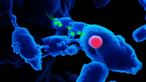
In the evolving landscape of cancer research, scientists are increasingly turning their attention to the mechanical properties of tissues as a critical factor in metastasis. Recent studies utilizing cellular force spectroscopy have uncovered compelling evidence that tissue stiffness plays a pivotal role in guiding cancer cell migration and invasion. This discovery challenges traditional views of metastasis as a purely biochemical process, highlighting how physical forces within the tumor microenvironment can dictate the spread of malignant cells.
The biomechanical dialogue between cancer cells and their surroundings has emerged as a key area of investigation. Researchers have observed that tumors often alter the extracellular matrix (ECM), creating stiffer regions that serve as pathways for migrating cells. These mechanical changes trigger a phenomenon known as durotaxis, where cancer cells preferentially move toward stiffer areas of tissue. The implications of this finding are profound, suggesting that the physical architecture of tissues may be just as important as chemical signals in determining metastatic patterns.
Advanced cellular force spectroscopy techniques have enabled scientists to measure these mechanical interactions with unprecedented precision. By quantifying the forces exerted by single cancer cells on their environment, researchers can map how stiffness gradients influence cell behavior. These measurements reveal that cancer cells not only respond to existing stiffness patterns but actively remodel their surroundings to create favorable mechanical conditions for migration. This two-way mechanical communication forms a self-reinforcing cycle that accelerates metastatic progression.
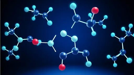
At the molecular level, the connection between tissue stiffness and cancer cell behavior appears to be mediated by mechanotransduction pathways. When cells encounter stiff substrates, mechanical forces are converted into biochemical signals through integrin receptors and cytoskeletal components. This triggers a cascade of events that promote invasive phenotypes, including the activation of focal adhesion kinase (FAK) and Rho-associated protein kinase (ROCK). These pathways ultimately lead to increased contractility and the formation of invasive protrusions that facilitate tissue penetration.
The clinical implications of these findings are becoming increasingly apparent. Diagnostic approaches that assess tissue stiffness, such as elastography, may provide valuable prognostic information about metastatic potential. Furthermore, therapeutic strategies targeting mechanotransduction pathways offer new possibilities for intervention. Drugs that disrupt the ability of cancer cells to sense or respond to mechanical cues could potentially block metastasis at its earliest stages, before malignant cells have the chance to spread to distant organs.
Emerging evidence suggests that the relationship between stiffness and metastasis may vary across different cancer types. While many carcinomas show increased invasiveness on stiffer substrates, some mesenchymal cancers demonstrate the opposite behavior. This complexity underscores the need for personalized approaches to cancer treatment that consider both the biochemical and biomechanical characteristics of individual tumors. Researchers are now working to develop comprehensive models that integrate these mechanical factors with traditional biomarkers.
Looking ahead, the field faces several important challenges. Developing standardized methods for measuring tissue mechanics in clinical settings remains a significant hurdle. Additionally, the dynamic nature of tissue stiffness during tumor progression adds another layer of complexity to therapeutic targeting. Despite these challenges, the growing understanding of how physical forces guide metastasis represents a paradigm shift in oncology, opening new avenues for early detection and intervention in cancer's deadly spread.
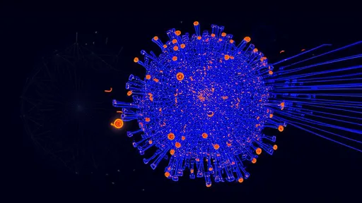
The integration of cellular force spectroscopy with other advanced technologies promises to further illuminate the mechanical underpinnings of cancer progression. Techniques such as atomic force microscopy and traction force microscopy are being combined with molecular biology approaches to create a more complete picture of how cells sense and respond to their mechanical environment. These multidisciplinary efforts are gradually revealing the intricate dance between physical forces and biological processes that governs metastatic behavior.
As research in this area continues to advance, it becomes increasingly clear that the fight against cancer metastasis must address both the chemical and physical dimensions of the disease. The stiffness of tissues, once considered merely a structural characteristic, is now recognized as an active participant in cancer progression. This realization not only deepens our understanding of metastasis but also provides fresh hope for developing more effective strategies to combat this deadly aspect of cancer.

By /Jul 28, 2025

By /Jul 28, 2025

By /Jul 28, 2025
By /Jul 28, 2025

By /Jul 28, 2025

By /Jul 28, 2025
By /Jul 28, 2025

By /Jul 28, 2025
By /Jul 28, 2025

By /Jul 28, 2025

By /Jul 28, 2025

By /Jul 28, 2025

By /Jul 28, 2025

By /Jul 28, 2025

By /Jul 28, 2025
By /Jul 28, 2025

By /Jul 28, 2025

By /Jul 28, 2025

By /Jul 28, 2025

By /Jul 28, 2025